Datei:An atlas of human anatomy for students and physicians (1919) (19722786094).jpg

Originaldatei(1.238 × 1.166 Pixel, Dateigröße: 450 KB, MIME-Typ: image/jpeg)

Diese Datei und die Informationen unter dem roten Trennstrich werden aus dem zentralen Medienarchiv Wikimedia Commons eingebunden.

Zur Beschreibungsseite auf Commons


Beschreibung

Beschreibung
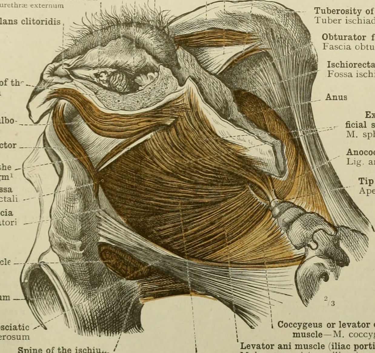
English:

Title: An atlas of human anatomy for students and physicians
Identifier: atlasofhumananat34told (find matches)
Year: 1919 (1910s)
Authors: Toldt, Carl, 1840-1920; Dalla Rosa, Alois, b. 1848; Paul, Eden, 1865-1944, tr; Toldt, Carl, 1840-1920. Anatomischer atlas für Studierende und Ärtze. English
Subjects: Anatomy
Publisher: New York, Rebman Company
Contributing Library: Columbia University Libraries
Digitizing Sponsor: Open Knowledge Commons

View Book Page: Book Viewer
About This Book: Catalog Entry
View All Images: All Images From Book
Click here to view book online to see this illustration in context in a browseable online version of this book.

Text Appearing Before Image:
MUSCLES OF THE PERINEU 529 Labium majus Labium majus pudend Vaginal orifice—Orificium vaginaei Labium minus, or nympha—Labium mmus, pudendi ; External orifice of the urethra Onticium Glans clitoridis Ischiocavemosus or erector clitoridis muscle 1 Superficial layer of the triangular ligament of the ; urethra^—Fascia diaphragmatis urogenitalis interior ' I Superficial transverse perineal muscle^ ' M trans\ersu3 perinei superhcialis Tuberosity of the ischium Tuber ischiadicum Wall of the vestibule of th vagina in section Sphincter vaginae or bulbo- cavemosus muscle Ischiocavemosus or erector clitoridis muscle Posterior border of the Obturator fascia Fascia obturatoria Ischiorectal fossa Fossa ischiorectalis 'urogenital diaphragm' \^ Ischiorectal fossa Fossa ischiorectal Obturator fascia Fascia obturator: Obturator internus muscle Posterior or great saorosciaiic '- ligament —Lig. sacrotuberosum Spine of the ischiu. Spina ischiadica
Text Appearing After Image:
External or super- ficial sphincter of the anus M. sphincter ani externus Anococcygeal ligament* Li^' anoroccygeum Tip of the coccyx" Apex ossis coccygis I Coccygeus or levator coccygis \ muscle—M. coccvKeus Levator ani muscle filiac portion)" M. levator ani (pars iliaca) Levator ani muscle (pubic portion) M. levator ani (pars pubica) Pelvic diaphragm Diaphragma pelvis Fig. 917.—The Muscles of the Perineal Region of the Female seen from the Left Side. Of the External Genital Organs, the Labium Majus and the Labium Minus or Nympha have been removed on the Left Side. Levator Ani and Coccygeus or Levator Coccygis Muscles, forming the Pelvic Diaphragm. cted poste in/er rs uf the superficial and deep layers of ihc triangular ligament of the • layer of the triangular ligament (see Appendix, note **). ethra (see Appendii « That is, the c note »),—Tk. » Known also as the anterior c 3 See note uh'c fiortion o/the levator ani mitscle, and by Savage the fiuSococcy^eus muscle. The posterior portion arises from the 'jjhite line 0/ the pelvic/ascia behind the obturator caruil and from the spine of the ischium ; it is called by Toldt the iliac portion 0/ the levator ani muscle, by Henle the ischiococcygcus muscle, and by Savage the obturatococcygeus muscle.—Tr. Musculi perinei—Muscles of the perineum. 67

Note About Images

Please note that these images are extracted from scanned page images that may have been digitally enhanced for readability - coloration and appearance of these illustrations may not perfectly resemble the original work.
Datum
Quelle

https://www.flickr.com/photos/internetarchivebookimages/19722786094/

Urheber Internet Archive Book Images
Genehmigung
(Weiternutzung dieser Datei)
At the time of upload, the image license was automatically confirmed using the Flickr API. For more information see Flickr API detail.
Volume
InfoField
v.3-4
Flickr tags
InfoField
Flickr posted date
InfoField
6. August 2015

Lizenz

Dieses Bild wurde von Flickrs The Commons übernommen. Die hochladende Organisation kann verschiedene Gründe für die Feststellung haben, dass keine bekannten Urheberrechtsbeschränkungen bestehen, wie z. B.:
  1. Gemeinfrei, da das Urheberrecht abgelaufen ist;
  2. Das Urheberrecht wurde aus anderen Gründen, wie z. B. der Nichteinhaltung von erforderlichen Formalitäten oder Bedingungen, gemeinfrei;
  3. Die Einrichtung besitzt das Urheberrecht, ist aber nicht an der Ausübung der Kontrolle interessiert; oder
  4. Die Institution verfügt über ausreichende Rechte, um anderen die uneingeschränkte Nutzung des Werkes zu gestatten.

Weitere Informationen findest du unter https://flickr.com/commons/usage/.


Bitte füge zusätzliche Lizenzvorlagen zu diesem Bild hinzu, wenn genauere Informationen zum Urheberrechts-Status ermittelt werden können. Siehe Commons:Licensing für weitere Informationen.
Diese Bilddatei wurde ursprünglich auf Flickr durch Internet Archive Book Images in https://flickr.com/photos/126377022@N07/19722786094 hochgeladen. Sie wurde am 17. September 2015 durch den FlickreviewR-Bot geprüft und die Lizenzierung der Datei unter den Bedingungen von No known copyright restrictions wurde bestätigt.

17. September 2015

Kurzbeschreibungen

Ergänze eine einzeilige Erklärung, was diese Datei darstellt.

In dieser Datei abgebildete Objekte

Motiv

image/jpeg

fc1b8b4b419e9b65dfeb66f8e8df9b9d6c3f022b

460.516 Byte

1.166 Pixel

1.238 Pixel

Dateiversionen

Klicke auf einen Zeitpunkt, um diese Version zu laden.

Version vomVorschaubildMaßeBenutzerKommentar
aktuell00:20, 18. Sep. 2015Vorschaubild der Version vom 00:20, 18. Sep. 20151.238 × 1.166 (450 KB)== {{int:filedesc}} == {{subst:chc}} {{information |description={{en|1=<br> '''Title''': An atlas of human anatomy for students and physicians<br> '''Identifier''': atlasofhumananat34told ([https://commons.wikimedia.org/w/index.php?title=Special%3ASear...

Keine Seiten verwenden diese Datei.